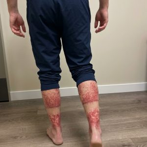
After the initial clinical assessment, the patient presented with a sudden onset of painful, erythematous lesions involving the face and neck, prompting immediate referral to dermatology for further evaluation. The abrupt appearance, tenderness, and distribution of the lesions raised concern for an acute inflammatory or hypersensitivity-driven process, leading the clinical team to initiate an urgent and structured diagnostic workup. One of the first clinical decisions was the discontinuation of a recently introduced medication due to a suspected temporal association with symptom onset. This step was taken as a precautionary measure while broader investigations were initiated to clarify the underlying cause of the cutaneous eruption.
A comprehensive diagnostic panel was ordered, including a skin biopsy to assess histopathological features, a complete blood count to evaluate inflammatory markers and hematologic abnormalities, and an expanded set of immunological and serological tests. These included antibody screening, lupus anticoagulant testing, and additional markers aimed at excluding connective tissue disease and infectious etiologies. The combination of acute skin involvement and systemic inflammatory suspicion required close monitoring, and systemic corticosteroid therapy was initiated to control symptoms while awaiting diagnostic confirmation.
Within 48 hours of corticosteroid administration, the patient demonstrated significant clinical improvement. The erythema began to fade, pain levels decreased, and lesion activity visibly subsided, indicating a strong response to anti-inflammatory treatment. This rapid improvement supported the working diagnosis of an immune-mediated inflammatory dermatosis and justified the continuation of corticosteroid therapy pending further results. Laboratory findings subsequently revealed leukocytosis with marked neutrophilia, a pattern commonly associated with acute inflammatory or reactive processes rather than chronic autoimmune disease. Immunological testing showed the presence of certain antibodies along with a positive lupus anticoagulant, while infectious serologies returned negative, creating a complex but non-specific diagnostic profile that required histological correlation.
Approximately twenty days after presentation, the skin biopsy results confirmed dense neutrophilic infiltration within the dermis without evidence of primary vasculitis, consistent with Sweet syndrome, also known as acute febrile neutrophilic dermatosis. This condition is classified among neutrophilic dermatoses and is characterized histologically by a prominent accumulation of mature neutrophils in the dermal layer. Clinically, it presents with the sudden eruption of painful erythematous papules, nodules, or plaques, often accompanied by systemic symptoms such as fever, malaise, and elevated inflammatory markers including leukocytosis and neutrophilia.
The pathophysiology of Sweet syndrome is not fully understood but is believed to involve immune dysregulation driven by cytokine-mediated activation of neutrophils. Elevated levels of inflammatory mediators such as interleukin-1, interleukin-6, and granulocyte colony-stimulating factor contribute to neutrophil proliferation and migration into cutaneous tissues. The condition may occur idiopathically or be triggered by infections, malignancies, or medications. Drug-induced cases have been reported with agents including antibiotics, antiepileptics, antihypertensives, vaccines, and hematopoietic growth factors, highlighting the importance of medication history in early evaluation.
In this case, the temporal relationship between symptom onset and recent medication exposure strongly suggested a drug-induced etiology, supporting early discontinuation of the suspected agent. Systemic corticosteroids remain the mainstay of treatment and typically produce rapid clinical improvement within days. The patient’s response within 48 hours was consistent with this expected therapeutic pattern, further reinforcing the diagnosis even before histological confirmation.
Differential diagnosis in such presentations includes urticaria, contact dermatitis, drug eruptions, and cutaneous lupus erythematosus, each of which can present with overlapping clinical features but differ in histopathology, progression, and systemic involvement. The integration of clinical findings, laboratory data, therapeutic response, and biopsy results was essential in distinguishing Sweet syndrome from these conditions. The final diagnosis allowed for continued targeted management and appropriate follow-up to monitor resolution and exclude any underlying systemic disease associations.